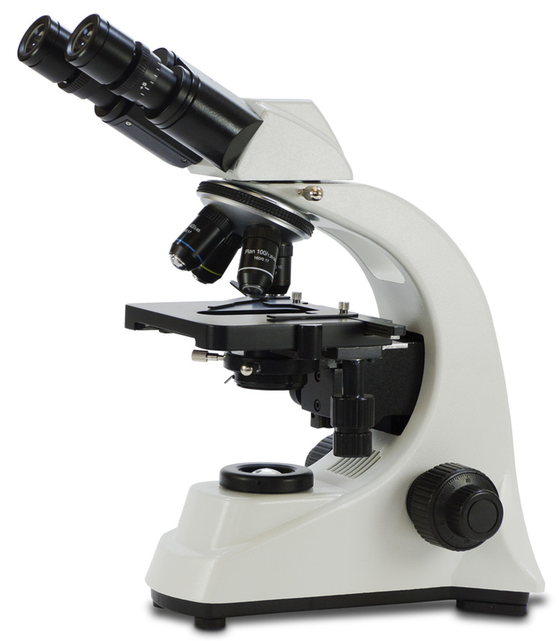
Experience laboratory-grade optical performance with the Laboratory Binocular Microscope featuring Plan Optics. Designed for comfortable extended viewing, this microscope combines superior plan objectives that deliver 95% flat-field clarity with an ergonomic Siedentopf binocular head. Perfect for biology labs, medical applications, or veterinary work where consistent visual observation is essential, this microscope provides exceptional edge-to-edge sharpness from 40x to 1000x magnification.
- Superior plan objectives deliver 95% of the field in sharp focus simultaneously—no constant refocusing needed when scanning specimens
- Comfortable Siedentopf binocular head with 360° rotation, dual diopter adjustment, and 55-75 mm interpupillary settings for customized viewing
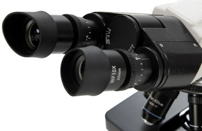
- Premium 20 mm wide-field eyepieces provide an expanded viewing area with long eye relief for comfortable extended use
- 1000x oil immersion capability for examining bacteria, blood cells, and fine cellular details with exceptional resolution
- Professional Abbe condenser with iris diaphragm and 20-watt halogen illumination for precise contrast control
- Backed by a lifetime warranty
 |
Laboratory Performance Without the Premium Price
This microscope delivers lab-grade plan optics and professional-quality mechanics at an accessible price. The flat-field correction and DIN standard objectives ensure accurate observations across the entire viewing field.
|
- Laboratory Binocular Microscope with Plan Optics
- Operating instructions
- Dust cover
- Spare halogen bulb
- Immersion oil
- Blue, green, and yellow contrast filters
- Eyepieces: Two 10x widefield high comfort eyepieces with 20 mm exit pupil distance and 22 mm super wide viewing field
- Head Design: Siedentopf binocular head, 30° inclined, rotates 360° and includes dual diopters and 55-75 mm interpupillary adjustment
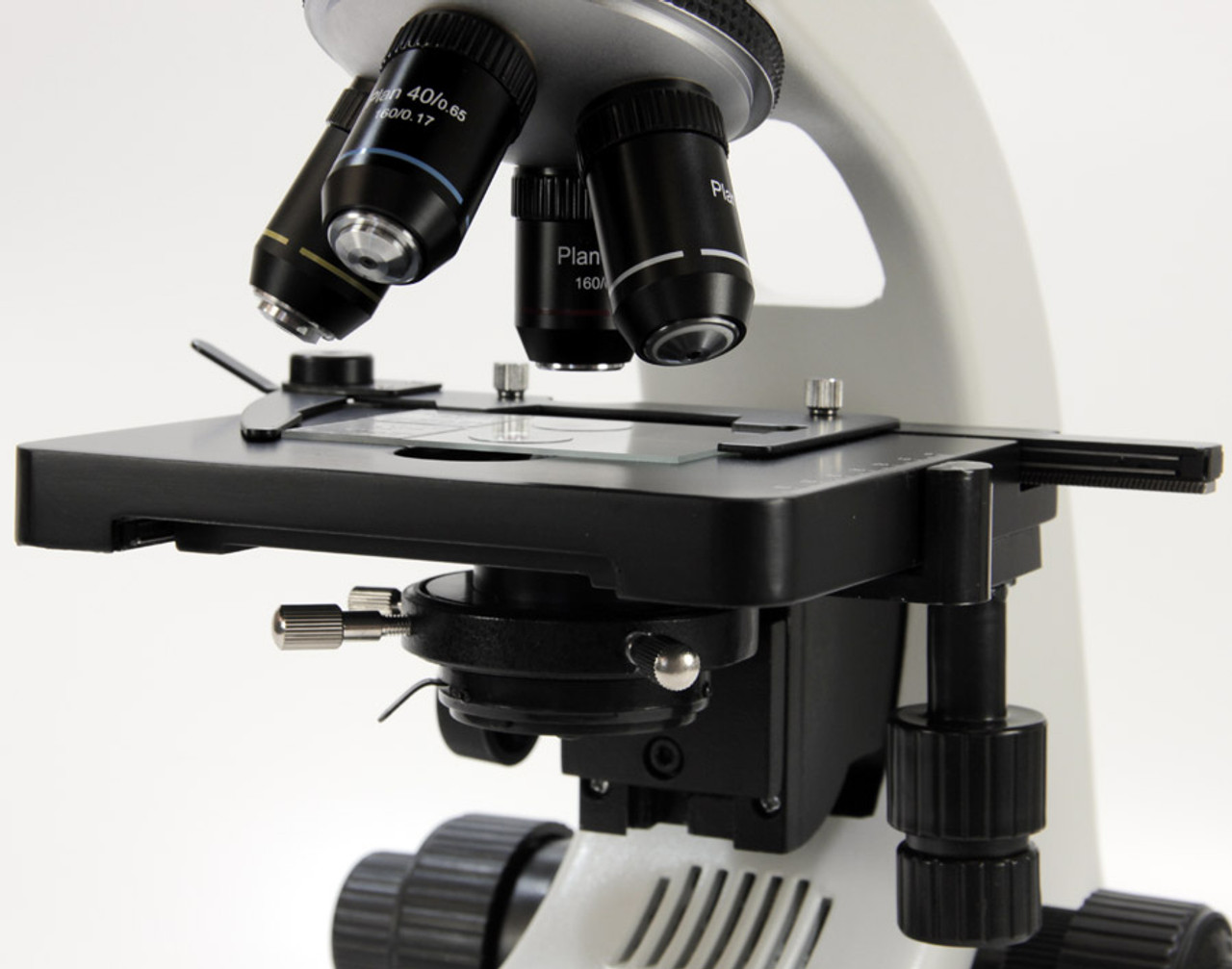
- Objectives: 4x (0.10 NA), 10x (0.25 NA), 40xR (0.65 NA) and 100xR (1.25 NA) superior plan objectives
- Objective Quality: DIN standard, parcentered, and parfocaled—observe virtually the entire viewing field in sharp focus at once
- Objective Features: 40xR objective is retractable; 100xR objective is oil retractable. 4-hole objective turret is ball-bearing mounted for smooth, precise changes in magnification
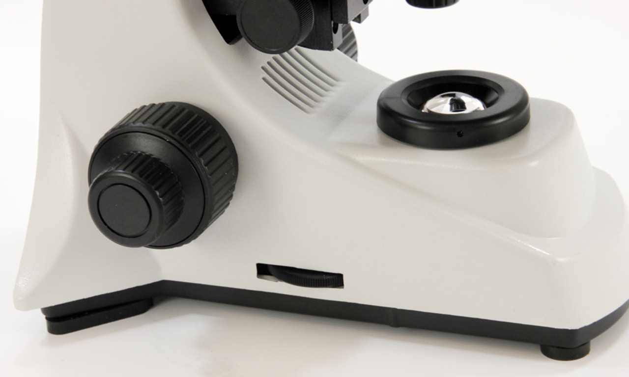
- Focus System: Smooth precision coaxial coarse and fine focusing with tension adjustment and 2 micron fine division. Focus tension adjustment eliminates stage drift
- Stage: Large 132 x 140 mm stage with stage stop and integrated ball-bearing double-layer 55 x 75 mm mechanical stage with x-y movement low-position coaxial controls
- Condenser: 1.25 N.A. Abbe condenser with rack-and-pinion focusing
- Illumination: Extra-bright 20-watt halogen illumination with variable intensity control for glare-free and uniform specimen illumination. Adjustable iris diaphragm for excellent contrast control
- Filters: Swing-out filter holder with blue, green, and yellow contrast filters
- Power Supply: High-quality 100-240 volt AC universal power supply
- Field of View: 5.0 mm with 4x, 2.0 mm with 10x, 0.5 mm with 40x and 0.2 mm with 100x objective
- Construction: Cast aluminum frame with chemical-resistant coating stands 16" tall, 8.5" wide, and 10" deep
- Also Available: Trinocular version with camera port, digital camera, and replacement light bulb
Laboratory Microscope for Professional Applications
The Laboratory Binocular Microscope with Plan Optics delivers exceptional optical performance for biology, medical, and veterinary laboratory work. The plan objectives use additional corrective lens elements to eliminate field curvature, keeping 95% of the viewing field in sharp focus simultaneously. This flat-field correction means you can scan across large tissue sections, blood smears, or microorganism samples without constantly refocusing—saving time and reducing eye strain during extended observation sessions.
The ergonomic Siedentopf binocular head provides comfortable viewing for hours of use. Unlike rigid binocular heads, the Siedentopf design allows interpupillary adjustment without changing the tube length, maintaining focus for all users. The 30° inclined viewing angle reduces neck strain, while the 360° rotation lets you share views with colleagues without moving the microscope. Dual diopter adjustment compensates for differences between your eyes, ensuring both see equally sharp images.
Professional Components for Reliable Results
This microscope includes professional-grade components found in much more expensive laboratory instruments. The ball-bearing mounted mechanical stage with low-position coaxial controls allows smooth, precise specimen movement while your hands remain in a natural position. The 1.25 N.A. Abbe condenser with iris diaphragm provides optimal specimen illumination and contrast control essential for examining stained tissue sections and microorganisms. The 20-watt halogen light source delivers bright, uniform illumination with variable intensity adjustment.
The four DIN standard plan objectives (4x, 10x, 40x retractable, and 100x oil immersion retractable) are parcentered and parfocaled, meaning specimens stay centered and nearly in focus when you change magnifications. The 100x oil immersion objective delivers the resolution needed to examine bacteria, blood cells, chromosomes, and other minute structures. DIN standardization ensures you can replace objectives if needed without compatibility concerns.
Choosing Between Binocular and Trinocular
This microscope is also available with a trinocular head that includes a third port for attaching a digital camera. The binocular version is ideal when your primary need is visual observation and direct examination of specimens. It offers comfortable viewing without the added complexity and cost of imaging capabilities. The simpler design also means all available light goes to the eyepieces for the brightest possible viewing experience. If you need to capture images for documentation, teaching, or sharing with colleagues, consider the trinocular version which allows simultaneous viewing and photography.
Frequently Asked Questions
- What makes plan optics different from standard achromatic objectives? Plan optics correct for field curvature, keeping 95% of the viewing area in sharp focus simultaneously, compared to standard achromatic objectives that keep only the center 65% focused. This eliminates constant refocusing when scanning specimens and ensures accurate photomicrography across the entire field. Plan objectives use additional corrective lens elements to deliver edge-to-edge clarity essential for examining large tissue sections and blood smears.
- Do I need oil immersion for 1000x magnification? Yes, oil immersion is essential at 1000x magnification. The immersion oil has a refractive index similar to glass, preventing light scattering between the slide and objective lens. Without oil, magnifications above 400x produce blurry images due to light loss. Oil immersion dramatically increases resolution, allowing you to clearly observe bacteria, individual blood cells, chromosomes during cell division, and other subcellular structures.
- Can I add a camera to a binocular microscope? While it's possible to attach a camera to one eyepiece of a binocular microscope using an adapter, this blocks that eyepiece during photography and can be awkward. For regular imaging needs, the trinocular version is more practical as it has a dedicated camera port that doesn't interfere with binocular viewing. However, if you only occasionally need to capture images and primarily use the microscope for direct observation, the binocular version with an occasional camera adapter may be sufficient.
- What applications is this laboratory binocular microscope best suited for? This microscope excels in biology, medical, and veterinary laboratories where consistent visual observation is the primary need. It's ideal for examining stained tissue sections, blood smears, microorganisms, parasitology samples, and cytology specimens. The plan optics and 1000x capability make it suitable for teaching environments, clinical diagnostics, quality control, and research applications where accurate observation of fine specimen details is essential but regular image capture is not required.